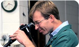
Endoscopy (bronchoscopy, esophagoscopy, peritoneoscopy and thoracoscopy)
 Endoscopy means looking inside and refers to looking inside the human body for medical reasons. Endoscopy, using a borescope, is also used in engineering and techical situations such as the inspection of nuclear fuel elements, aircraft or engines where direct line-of-sight observation is not feasible. The instrument used is called an endoscope..
ENDOSCOPY examination of the inside of an organ or body cavity using a fiberoptic instrument. The report should describe the condition of the organ with reference to swelling, blockage, lesions, growths, and other abnormalities.
Key words/possible involvement: mass or lesion visualized in the opening, or if a biopsy via the endoscope yields a diagnosis of malignancy, fixation; stricture, polyp, adenoma, lesion, neoplasm, malignancy. Other words/no involvement: no abnormalities visualized during the examination , no strictures or foreign bodies; inflammatory process, foreign bodies, abscess, infectious process, or other benign conditions. Key information: largest size of tumor, gross description of tumor, presence of multiple tumors, degree of induration of ureteric wall, extension outside of organ (kidney or ureter).
BRONCHOSCOPY endoscopic visualization of the trachea and mainstem and lobar bronchi to evaluate invasion from lung or from esophagus, using a lighted tube inserted into the lungs through the mouth. Key words/possible involvement: mass or lesion visualized in the bronchial tree, or if a biopsy via the bronchoscope yields a diagnosis of malignancy.
Other words/no involvement: no abnormalities visualized during the examination
COLONOSCOPY examination of the large intestine using a fiberoptic instrument. The report should describe the condition of the colon in the cecum, ascending, hepatic flexure, transverse, splenic flexure, and descending portions of the colon, in addition to the sigmoid and rectum. Colonoscopy generally examines the colon to a level of 60 cm or higher. Key words/possible involvement: stricture, polyps, villous adenoma, lesion, neoplasm, malignancy. Other words/no involvement: diverticulosis, megacolon, ulcerative colitis, Crohn's disease, inflammatory process, foreign bodies, abscess, or infectious process, or other benign conditions. Words indicating unsatisfactory procedure: not satisfactory due to residual fecal material in the colon or incomplete preparation of the colon.
COLPOSCOPY examination of the vagina and cervix through a colposcope, an instrument containing a magnifying lens that is inserted into the vagina.
Key words/possible involvement: lesion, tumor, leukoplakia, whitish areas of epithelium, gray area, area of discoloration, bleeding, mosaic pattern, mosaic staining, Toluidine staining, Iodine staining, irregular blood vessels, infiltrated patches, atypical epithelium, abnormal epithelium, suspicious lesion, neoplasm, malignancy, ulceration, exophytic lesion, infiltration.
Other words/no involvement: no abnormalities visualized during the examination.
CYSTOSCOPY examination of the bladder using a fiberoptic instrument. Usually not performed for colon tumors. May be performed for a fixed or highly invasive rectal tumor.
Key words/possible involvement: bullous edema, lesion, tumor invasion, extrinsic mass, tumor infiltration, invasion of bladder mucosa, extension of tumor into bladder wall.
Other words/no involvement: if there is no reference to tumor or abnormality in the bladder.
CYSTOURETHROSCOPY examination of the bladder and urethra using a fiberoptic instrument.
DUODENOSCOPY endoscopic visualization of the upper portion of the small intestine (duodenum).
ERCP (ENDOSCOPIC RETROGRADE CHOLANGIOPANCREATOGRAPHY) Evaluation of the gallbladder and pancreas using contrast material instilled in the duodenum or ampulla of Vater via an endoscope. Key words/possible involvement: hypervascularity, stricture, extrinsic mass, lesion, neoplasm, malignancy, opacification, nonvisualization, stones, stenosis.
Other words/no involvement: if there is no specific reference to visible abnormality in the organ; inflammatory process, foreign bodies, or other benign conditions.
ESOPHAGOGASTRODUODENOSCOPY Also called EGD. Consists of visualization of esophagus, stomach and small intestine (duodenum) as part of a single procedure.
ESOPHAGOSCOPY endoscopic visualization of the esophagus to evaluate invasion from a lung or stomach tumor.
GASTROSCOPY endoscopic visualization of the stomach to evaluation invasion from other organs.
HYSTEROSCOPY examination of the uterus using a fiberoptic instrument. Key words/possible involvement: tumor, leukoplakia, whitish areas of epithelium, irregular blood vessels, infiltrated patches, atypical epithelium, abnormal epithelium, suspicious lesion, neoplasm, malignancy. Other words/no involvement: no abnormalities visualized during the examination.
LAPAROSCOPY examination of the inside of the abdomen using a fiberoptic instrument. The report should describe the condition of organs in the abdomen with reference to swelling, blockage, lesions, growths, and other abnormalities. Key words/possible involvement: mass, lesion, abnormal lymph nodes, seeding, salt and pepper, talcum powder appearance, nodules, caking, implants, encasement, frozen pelvis, matted organs.
Other words/no involvement: no abnormalities visualized during the examination; adhesions.
LARYNGOSCOPY endoscopic visualization of the larynx to evaluate for a head and neck primary tumor; to determine a cause for vocal cord paralysis other than recurrent laryngeal nerve paralysis due to involvement by lung cancer; or to determine invasion from esophagus.
MEDIASTINOSCOPY an invasive endoscopic procedure to biopsy the lymph nodes in the mediastinum by means of a bronchoscope inserted through an incision in the base of the neck. Key words/possible involvement: mass, lesion, or abnormal lymph nodes visualized in the mediastinum, or if a biopsy of the mediastinum yields a diagnosis of malignancy.
Other words/no involvement: no abnormalities visualized during the examination.
NASOPHARYNGOSCOPY endoscopic visualization of the nasopharynx and pharynx to evaluate region for primary or secondary malignancy.
PERITONEOSCOPY endoscopic examination of the peritoneum. Key words/possible involvement: mass, lesion, abnormal lymph nodes, nodules, encasement, frozen pelvis, matted organs.
Other words/no involvement: no abnormalities visualized during the examination; adhesions.
PROCTOSIGMOIDOSCOPY examination of the lower portion of the large intestine (sigmoid and rectum) using a fiberoptic instrument. Also called: proctoscopy, sigmoidoscopy. Proctosigmoidoscopy generally describes the condition of the lower colon to a level of 12 inches or 31 cm., or to 60 cm, depending on the instrument used. Key words/possible involvement: stricture, polyps, villous adenoma, lesion, neoplasm, malignancy, invasion of rectal mucosa, extension of tumor into rectal wall.
Other words/no involvement: diverticulosis, megacolon, ulcerative colitis, Crohn's disease, inflammatory process, foreign bodies, abscess, or infectious process, or other benign conditions. Words indicating unsatisfactory procedure: not satisfactory due to residual fecal material in the colon or incomplete preparation of the colon.
SIGMOIDOSCOPY examination of the lower portion of the large intestine (sigmoid and rectum) using a fiberoptic instrument. Sigmoidoscopy generally describes the condition of the lower colon to a level of 12 inches or 31 cm., or to 60 cm, depending on the instrument used. Also called: proctoscopy, proctosigmoidoscopy. Key words/possible involvement: stricture, polyps, villous adenoma, lesion, neoplasm, malignancy.
Other words/no involvement: diverticulosis, megacolon, ulcerative colitis, Crohn's disease, inflammatory process, foreign bodies, abscess, or infectious process, or other benign conditions. Words indicating unsatisfactory procedure: not satisfactory due to residual fecal material in the colon or incomplete preparation of the colon.
THORACOSCOPY endoscopic visualization of the thoracic cavity. Also called pleural endoscopy.
TRIPLE ENDOSCOPY (also called panendoscopy) combination procedure that examines the trachea, larynx, pharynx and esophagus via endoscopic visualization; used to investigate all mucosal surfaces of the upper respiratory tract for original or subsequent primaries.
URETEROSCOPY examination of the renal pelvis and ureters using a fiberoptic instrument (usually performed under general anesthesia).<< back to "Paediatric Surgery"
 Apollo Hospital
 Fortis Hospital
 Artemis Hospital
 Medanta Hospital


 Jaslok Hospital
 Lilavati Hospital

 Global Hospitals
 Jupiter Hospital


